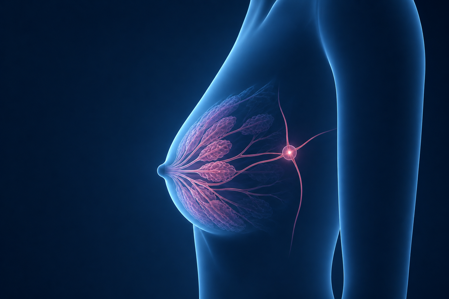
A patient-friendly guide to how mammograms, ultrasound, and breast MRI help find breast cancer, why additional imaging may be needed, and what a callback usually means.
A mammogram is a low-dose X-ray picture of the breast. It helps radiologists look for changes that may be too small to feel, including tiny calcifications, masses, areas of distortion, or a new change compared with prior mammograms.
On a mammogram, fatty tissue usually looks darker, while dense breast tissue and many breast findings look white. Radiologists carefully look for areas that stand out from the surrounding tissue or have changed over time.
During the exam, the breast is gently compressed between two plates. Compression can be uncomfortable, but it is important because it spreads the tissue out, improves image quality, lowers the radiation dose, and helps reduce blur. Most screening mammograms include two views of each breast.
Many centers now use 3D mammography, also called tomosynthesis. This creates thin image slices through the breast, allowing the radiologist to look through the tissue layer by layer. This can be especially helpful when breast tissue overlaps or when the breasts are dense.
Early
detection often gives patients more treatment options
3D
mammography helps reduce the effect of overlapping tissue
Tiny
calcifications can sometimes be seen before a lump forms
Compare
prior mammograms are very helpful for spotting change
Screening vs. diagnostic mammogram: A screening mammogram is a routine exam when there is no specific breast concern. A diagnostic mammogram is done when there is a symptom, a callback from screening, or a finding that needs a closer look. Diagnostic imaging may include extra mammogram views and ultrasound.
What Cancer Can Look Like on Mammography
Breast cancer does not have one single appearance on imaging. Sometimes it looks like a mass. Sometimes it appears as tiny calcifications. Other times it may show up as pulling, distortion, or an area that looks different from the rest of the breast.
It is important to remember that many findings seen on mammograms are not cancer. Imaging helps radiologists decide whether something looks benign, probably benign, or suspicious enough that more evaluation is needed.
How Mammogram Images Work
Different tissues show up in different shades. Fat is usually darker. Dense tissue, masses, and calcifications often appear lighter or white. This is one reason dense tissue can make mammograms harder to read.
■ Black Air
■ Dark Gray Fat
■ Medium Gray Fibrous tissue
■ Light Gray Glandular tissue
■ Off-White Dense tissue
■ Bright White Calcification / Mass
Spiculated Mass
A mass with irregular, spiky edges can be concerning because it may suggest the finding is growing into surrounding tissue. This type of appearance usually needs additional evaluation and often biopsy.
Irregular Mass
An irregular or poorly defined mass is more concerning than a smooth, round mass. Ultrasound is often used to better understand whether the area is solid, cystic, or needs biopsy.
Calcifications
Calcifications are tiny calcium deposits that look like small white specks. Most are benign, but certain shapes or patterns can be associated with early breast cancer or DCIS and may require biopsy.
Architectural Distortion
This means the normal breast tissue pattern looks pulled, twisted, or distorted without a clear mass. It can be subtle, but it is an important finding that usually needs a closer look.
Asymmetry
An asymmetry means one area of the breast looks different from the same area on the other side or from prior mammograms. Many asymmetries are benign, but a new or developing asymmetry may need more imaging.
Round or Oval Mass
Smooth, round, or oval masses are often benign, such as cysts or fibroadenomas. Ultrasound can be very helpful in telling whether a mass is fluid-filled or solid.
About calcifications: Calcifications are common, and most are not cancer. Radiologists pay close attention to their shape, size, and pattern. The pattern is what determines whether they can be safely ignored, followed, or biopsied.
Why Some Cancers Are Hard to See
Mammography is an excellent screening tool, but no imaging test finds every cancer. Some cancers are difficult to see because of dense breast tissue, subtle growth patterns, or because they grow quickly between screening exams.
This does not mean mammograms are not useful. Regular screening remains one of the most important tools for finding breast cancer early. It does mean that symptoms still matter, even if a recent mammogram was normal.
🔍 Dense Tissue Can Hide Findings
Dense breast tissue appears white on a mammogram, and many cancers also appear white. When white tissue overlaps a white mass, the finding can be harder to see. This is called masking.
For patients with dense breasts, additional imaging such as ultrasound or MRI may sometimes be recommended depending on individual risk.
🧬 Some Cancers Grow Quickly
Some breast cancers grow faster than others. A cancer may not have been visible on a prior mammogram but may become detectable months later. These are sometimes called interval cancers.
This is one reason it is important to report any new lump, skin change, nipple discharge, or persistent focal breast pain, even between routine mammograms.
📸 Some Cancers Do Not Form a Clear Lump
Invasive lobular carcinoma can grow in a more subtle, spread-out pattern. Instead of forming a round mass, it may cause only mild asymmetry or distortion, which can be difficult to detect on mammography.
When lobular cancer is diagnosed, MRI may be used to better understand the full extent of disease.
⚠️ Image Quality and Comparison Matter
Small findings can be subtle. Good positioning, clear images, and comparison with prior mammograms all help radiologists detect important changes.
→Prior mammograms help show whether something is new or stable
→Extra views may be needed if tissue overlaps
→Ultrasound or MRI may be recommended when mammography alone is limited
Know your own breasts: A normal mammogram is reassuring, but it does not replace paying attention to new symptoms. If you feel a new lump or notice skin changes, nipple discharge, or a persistent new area of concern, contact your healthcare provider.
Dense Breasts and Cancer Detection
Breast density describes how much fibroglandular tissue is present compared with fatty tissue on a mammogram. Dense breasts are very common and are not abnormal.
Density matters for two main reasons. First, dense tissue can make mammograms harder to read because both dense tissue and cancers can appear white. Second, dense breasts are associated with a modestly higher risk of breast cancer compared with mostly fatty breasts.
BI-RADS A
Almost Entirely Fatty
Mostly fatty tissue. Mammograms are usually easier to interpret.
BI-RADS B
Scattered Fibroglandular
Some scattered areas of dense tissue. Mammograms still work well overall.
BI-RADS C
Heterogeneously Dense
Many areas of dense tissue. Some small cancers may be harder to see.
BI-RADS D
Extremely Dense
Most of the breast appears dense. Mammography can be more limited.
If you have dense breasts, your mammogram is still important. Depending on your personal risk factors, your doctor may also discuss supplemental screening, such as ultrasound, breast MRI, or contrast-enhanced mammography.
💬 What to Do If You Have Dense Breasts
→Continue recommended mammogram screening
→Ask whether 3D mammography is available or appropriate for you
→Discuss whether supplemental ultrasound or MRI makes sense based on your risk
→Tell your doctor about family history, prior biopsies, or genetic risk factors
MRI vs Mammogram vs Ultrasound
Each breast imaging test answers a different question. One test is not automatically “better” than another in every situation. The best test depends on why imaging is being done, what was seen on prior exams, your breast density, and your personal risk level.
Feature
Mammogram
Ultrasound
Breast MRI
CEM *
Technology
Low-dose X-ray
Sound waves
Magnetic field + contrast dye
Mammogram + iodine contrast
Best used for
Routine screening and calcifications
Evaluating a specific lump or mammogram finding
High-risk screening and mapping known cancer
Finding cancers that enhance with contrast
Strength
Excellent at finding calcifications and subtle changes over time
Can tell cyst from solid mass and guide biopsy
Very sensitive, especially in dense breasts and high-risk patients
Combines mammogram detail with contrast information
Limitation
Dense tissue can hide findings
Not usually used alone for routine screening
Can find many benign areas that need workup
Not available everywhere and uses contrast
Uses contrast / dye
No
No
Yes
Yes
Radiation
Low-dose radiation
None
None
Low-dose radiation
Common role
Screening and first-line breast imaging
Problem-solving after mammogram or for a palpable lump
High-risk screening, pre-surgical planning, and extent of disease
Alternative or supplemental contrast-based breast imaging
* CEM = Contrast-Enhanced Mammography.
🏥 Mammogram - The Foundation
Mammography is the main screening test for breast cancer. It is widely available, quick, and very good at finding calcifications and changes compared with prior exams.
🔊 Ultrasound - The Problem-Solver
Ultrasound is often used when a specific area needs more evaluation. It can show whether a lump is a simple cyst, a solid mass, or something that needs biopsy.
🦸 MRI - The Most Sensitive
Breast MRI is very sensitive and can be especially helpful for high-risk screening, dense breasts, invasive lobular cancer, or planning treatment after cancer is diagnosed.
Why Additional Imaging Is Sometimes Needed
Being asked to return for more imaging can feel stressful. In many cases, it simply means the radiologist needs a clearer or more targeted look before making a final assessment.
Breast imaging is often a step-by-step process. A screening mammogram may identify an area that needs extra views. Ultrasound may then help determine whether that area is a cyst, a solid mass, normal tissue, or something that should be biopsied.
📈 Measuring the Full Extent
After breast cancer is diagnosed, MRI or contrast-enhanced imaging may be used to see how large the cancer is and whether there are any additional areas that need attention before surgery.
🔊 Clarifying a Finding
Ultrasound is commonly used to look more closely at a mass or asymmetry. It can often tell whether something is a simple cyst, which is benign, or a solid finding that needs closer evaluation.
🧬 Higher-Risk Screening
Patients with a higher lifetime risk of breast cancer may be advised to have breast MRI in addition to mammography. This decision depends on family history, genetics, prior biopsies, and other risk factors.
📊 Monitoring Treatment Response
When chemotherapy is given before surgery, imaging may be repeated to see how well the cancer is responding and to help plan the next step in treatment.
Biopsy gives the final answer: Imaging can show whether something looks benign or suspicious, but it usually cannot prove cancer with 100% certainty. When needed, biopsy allows a pathologist to examine tissue under a microscope.
Why Callbacks Happen
Getting called back after a screening mammogram can be frightening. I want you to know that a callback does not mean you have breast cancer. It means the radiologist saw something that needs a closer look before a final answer can be given.
Most callbacks turn out to be benign. Common reasons include overlapping tissue, cysts, calcifications, dense tissue, or an area that looks different compared with prior mammograms.
Common
callbacks happen frequently after screening mammograms
Usually
most callbacks do not result in a cancer diagnosis
Often
additional views or ultrasound provide the answer
More
common on a first mammogram without priors to compare
Common Reasons for a Callback
1
An Area Needs a Closer Look
The radiologist may see a possible mass, calcifications, asymmetry, or distortion. Extra mammogram views and ultrasound can often clarify whether this is real or simply overlapping tissue.
2
Dense Breast Tissue
Dense tissue can make mammograms harder to read. Sometimes additional imaging is recommended because dense tissue can hide small findings.
3
Image Quality or Positioning
Occasionally, images need to be repeated because not enough tissue was included, there was motion, or something on the skin created an artifact. This type of callback does not necessarily mean there is a breast problem.
4
A Change From Prior Mammograms
A new finding is more important than one that has been stable for years. This is why comparison with prior mammograms is so valuable.
5
Your First Mammogram
Callbacks are more common when there are no prior studies for comparison. What looks unusual on a first mammogram may later prove to be your normal breast tissue pattern.
What Happens After a Callback
A
Diagnostic Mammogram
Additional mammogram views focus on the area in question. Many findings disappear or become clearly benign with these extra views.
B
Ultrasound
Ultrasound may be used to look at a specific area. It can often tell whether something is a benign cyst or a solid finding that needs more evaluation.
C
Short-Term Follow-Up
If a finding is probably benign, follow-up imaging may be recommended in about 6 months to make sure it stays stable.
D
Biopsy
If a finding looks suspicious, biopsy may be recommended. A biopsy does not mean you have cancer; it means tissue is needed so a pathologist can give a definite answer.
📋 BI-RADS - Your Mammogram's Report Category
Most breast imaging reports include a BI-RADS category. This number helps communicate how concerned the radiologist is and what should happen next:
→BI-RADS 0: More imaging is needed before a final answer can be given
→BI-RADS 1: Negative - no suspicious finding
→BI-RADS 2: Benign - a non-cancerous finding
→BI-RADS 3: Probably benign - short-term follow-up is recommended
→BI-RADS 4: Suspicious - biopsy is usually recommended
→BI-RADS 5: Highly suspicious - biopsy is strongly recommended
→BI-RADS 6: Known biopsy-proven cancer
Have a breast imaging report? I can help translate the medical language into a clear, patient-friendly explanation so you can better understand what your report is saying.
Medical Disclaimer: This content is for patient educational purposes only and is not a substitute for professional medical advice, diagnosis, or treatment. Always seek the guidance of your physician or qualified health provider regarding your medical condition. Content is physician-reviewed and curated for patient understanding.