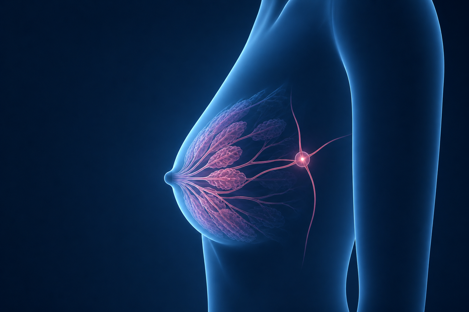
Not all breast cancers are the same. Understanding which type you have - and what makes it unique - is the first step toward informed, confident conversations with your care team.
The earliest form of breast cancer - abnormal cells fully contained inside a milk duct.
Non-Invasive · Stage 0
~20%
of new breast cancer diagnoses
Stage 0
earliest possible cancer stage
Highly Curable
with appropriate treatment
Mammogram
most cases found on screening
DCIS stands for Ductal Carcinoma In Situ - a Latin phrase meaning "in place." It is the earliest form of breast cancer, in which abnormal cells have developed inside one of the breast's milk ducts but have not broken through the duct wall into the surrounding breast tissue. Because the cells haven't invaded, DCIS cannot spread to lymph nodes or other organs at this stage.
DCIS is sometimes called a "pre-cancer" or Stage 0 breast cancer. While it is not yet life-threatening, it is important to treat it because, if left alone, some cases of DCIS can eventually progress into invasive breast cancer. The challenge is that doctors cannot always predict which DCIS cases will progress and which won't - which is why treatment is generally recommended.
How is DCIS found? Most DCIS cases are detected on a screening mammogram - they often show up as tiny calcium deposits (called calcifications) or a subtle shadow. DCIS usually causes no pain and no lump you can feel, which is why regular mammograms are so important.
High grade: cells look very abnormal; faster growing
Grade helps guide treatment intensity
Treatment Options
1
Surgery (First Step)
Breast-conserving surgery (lumpectomy) removes the DCIS area and a margin of normal tissue. If DCIS is widespread or large, mastectomy (removal of the whole breast) may be recommended. A small clip is often placed to mark the area during biopsy for the surgeon to find later.
2
Radiation Therapy (Often After Lumpectomy)
Radiation is typically given after breast-conserving surgery. Research shows it cuts the risk of DCIS returning in half. It is not needed after mastectomy. In some low-risk, older patients, radiation may be omitted after careful discussion.
3
Hormone Therapy (If Estrogen Receptor Positive)
If the DCIS tests positive for estrogen receptors (ER+), hormone-blocking medications such as tamoxifen (for pre-menopausal women) or anastrozole (for post-menopausal women) may be recommended for 5 years to reduce the risk of recurrence or a new breast cancer.
●
Invasive Ductal Carcinoma (IDC)
The most common breast cancer - begins in a milk duct and spreads into surrounding breast tissue.
Invasive · Most Common Type
70–80%
of all breast cancers are IDC
>99%
5-yr survival when localized (no spread)
87%
5-yr survival with regional spread
All ages
most common male breast cancer too
Invasive ductal carcinoma (IDC) - also called infiltrating ductal carcinoma - starts in the cells lining a breast milk duct and then breaks through the duct wall into the surrounding breast tissue. From there, it has the potential to spread to nearby lymph nodes and, if untreated, to distant organs via the lymphatic system or bloodstream.
IDC is the most common form of breast cancer in both women and men. The word "invasive" describes that the cells have left their original location - it does not mean the cancer has necessarily spread beyond the breast. Many IDC cases are caught at an early stage when they are very treatable. Treatment and prognosis depend on the tumor size, grade, hormone receptor status, HER2 status, and lymph node involvement.
🔍 Signs & Symptoms
A new lump or thickening in the breast
Change in breast size or shape
Nipple discharge or pain
Skin changes (dimpling, puckering)
Often found on mammogram before symptoms
🧬 Subtypes of IDC
ER/PR-positive IDC: hormone receptor positive - often responds to hormone therapy
HER2-positive IDC: overexpresses HER2 protein - responds to targeted therapy
Triple-negative IDC: no receptors - treated with chemotherapy
Rare subtypes: mucinous, medullary, tubular (see Less Common section)
💉 Treatment Overview
Surgery: lumpectomy or mastectomy depending on size/stage
Radiation: often after breast-conserving surgery
Chemotherapy: may be given before or after surgery
Hormone therapy: for ER/PR-positive IDC
Targeted therapy: for HER2-positive IDC
📋 What Your Pathology Report Will Tell You About IDC
After your biopsy or surgery, your pathology report will describe the IDC's grade (1–3, how abnormal cells look), size, margin status (whether cancer-free tissue surrounds the tumor), lymph node involvement, and receptor status (ER, PR, HER2). Together, these factors determine your stage and guide your treatment plan.
〰️
Invasive Lobular Carcinoma (ILC)
Starts in the milk-producing lobules - often harder to detect because it grows in a diffuse, sheet-like pattern rather than a lump.
Invasive · 2nd Most Common
10–15%
of invasive breast cancers
Age 55+
about 2/3 diagnosed at this age
E-cadherin
missing protein explains its growth pattern
MRI
often needed for accurate extent assessment
Invasive lobular carcinoma (ILC) begins in the lobules - the milk-producing glands at the end of the breast ducts - and then grows into the surrounding breast tissue. ILC is the second most common type of invasive breast cancer. What makes ILC unique is how it grows: ILC cells typically lack a protein called E-cadherin that normally keeps cells together. Without it, ILC cells spread in single-file lines through fatty breast tissue rather than forming a compact mass.
This diffuse, sheet-like growth pattern makes ILC uniquely challenging: it can be difficult to see on a standard mammogram and may not form a palpable lump. Instead, you might notice a vague area of thickening or fullness. Because of this, ILC is often larger at the time of diagnosis than IDC. Breast MRI or contrast-enhanced mammography (CEM) are significantly more accurate at detecting the full extent of ILC.
Imaging tip: If your biopsy confirms ILC, your doctor will likely recommend a breast MRI or contrast-enhanced mammogram to accurately measure the full extent of the cancer before surgery. Standard mammograms and ultrasound can underestimate how large ILC is. MRI helps surgeons plan the most effective operation.
🔍 Signs & Symptoms
Vague area of fullness or thickening (not a distinct lump)
Nipple turning inward (inversion)
Change in breast skin texture
Asymmetry between breasts
Some cases cause no symptoms at all
📸 Why Imaging Is Tricky
No E-cadherin protein → cells spread without forming a compact mass
Mammogram may only show subtle architectural distortion
Higher false-negative rate on standard mammogram vs. IDC
Breast MRI or CEM recommended for full staging
💉 Treatment Overview
Surgery: lumpectomy or mastectomy (guided by MRI findings)
ILC is usually ER/PR-positive → hormone therapy is key
Radiation often used after breast-conserving surgery
Chemotherapy when higher risk or node-positive
⚡
Triple-Negative Breast Cancer (TNBC)
Tests negative for all three main receptors - ER, PR, and HER2 - making it different to treat but often very responsive to chemotherapy.
Invasive · No Targeted Receptors
10–15%
of all breast cancers are triple-negative
Faster Growth
tends to grow and spread more quickly
Chemo Effective
often responds very well to chemotherapy
BRCA Link
associated with BRCA1/2 mutations
Triple-negative breast cancer (TNBC) gets its name from a laboratory test result: the cancer cells test negative for estrogen receptors (ER−), progesterone receptors (PR−), and HER2 (HER2−). These three receptors are the most common "drivers" that fuel breast cancer growth. Because TNBC lacks all three, it cannot be treated with hormone therapy (like tamoxifen) or HER2-targeted therapy (like trastuzumab).
TNBC tends to be more aggressive - it grows faster, is more likely to spread before diagnosis, and is more likely to recur than hormone-positive cancers. However, there is an important upside: TNBC often responds exceptionally well to chemotherapy. When treated with chemotherapy before surgery, many patients achieve a complete pathological response (no cancer remaining in the tissue) - a strong predictor of long-term cure. Newer treatments including immunotherapy and PARP inhibitors are also improving outcomes significantly.
🧬 TNBC and BRCA Mutations
Triple-negative breast cancer is more common in people who carry BRCA1 gene mutations. If you are diagnosed with TNBC - especially at a younger age - your doctor may recommend genetic testing. If a BRCA1 or BRCA2 mutation is found, it opens the door to additional targeted treatments called PARP inhibitors (such as olaparib), which specifically target cancer cells with BRCA-related DNA repair problems.
A BRCA mutation result also has implications for your family members, as it is inherited. Your care team can connect you with a genetic counselor.
How TNBC Is Treated
1
Chemotherapy (Often Before Surgery)
For most TNBC cases, chemotherapy is given before surgery (called neoadjuvant chemotherapy) to shrink the tumor. This approach also allows doctors to see how well the cancer responds. A combination of anthracyclines (e.g., doxorubicin) and taxanes (e.g., paclitaxel) is commonly used, sometimes alongside immunotherapy (e.g., pembrolizumab/Keytruda).
2
Surgery
Lumpectomy or mastectomy depending on tumor size, response to chemo, and patient preference. Sentinel lymph node biopsy or axillary dissection assesses lymph node involvement.
3
Post-Surgery Treatment
Radiation therapy is usually given after breast-conserving surgery. If cancer remains after chemotherapy, additional chemotherapy (capecitabine) may be recommended. For BRCA-positive patients, PARP inhibitors or antibody-drug conjugates (e.g., sacituzumab govitecan) may be options.
✦
HER2-Positive Breast Cancer
Driven by an overabundance of a growth protein called HER2 - once considered very aggressive, now one of the most treatable types thanks to targeted therapies.
Invasive · Targeted Therapy Available
~20%
of breast cancers are HER2-positive
>90%
5-yr survival when caught early with targeted therapy
−50%
reduction in recurrence risk with trastuzumab + chemo
Herceptin
trastuzumab - the landmark HER2-targeted drug
HER2 stands for Human Epidermal growth factor Receptor 2 - a protein found on the surface of all breast cells that helps control how cells grow and divide. In HER2-positive breast cancer, the cancer cells have too many copies of the HER2 gene (called amplification), which causes them to produce an excess of HER2 receptors. This flood of receptors sends constant "grow and divide" signals to the cell, making the cancer more aggressive and faster-growing.
For decades, HER2-positive breast cancer was considered one of the worst prognoses. That changed dramatically in the 1990s and 2000s with the development of targeted therapies specifically designed to block the HER2 receptor. Trastuzumab (Herceptin) - the first HER2-targeted drug - combined with chemotherapy reduces the risk of recurrence by 50% and improves overall survival by 30%. Today, survival rates for early-stage HER2-positive breast cancer exceed 90%, matching outcomes for hormone-positive disease.
What does HER2 testing mean? Your pathology report will show HER2 results as positive (3+), negative (0 or 1+), or equivocal (2+). An equivocal result is re-tested using a method called ISH (in-situ hybridization) to count gene copies. HER2 status is essential to know - it determines whether you are eligible for HER2-targeted treatments.
HER2-Targeted Therapies
💊 Monoclonal Antibodies
Trastuzumab (Herceptin): attaches to HER2 receptors and blocks growth signals; the cornerstone of HER2 treatment
Pertuzumab (Perjeta): blocks a different part of HER2; often combined with trastuzumab
Margetuximab (Margenza): used for advanced breast cancer with prior treatment
Trastuzumab deruxtecan (Enhertu): highly effective newer drug; also used in HER2-low cancers
💉 Full Treatment Plan
Chemotherapy + trastuzumab (± pertuzumab) - often before surgery
Surgery: lumpectomy or mastectomy based on response and stage
Radiation after breast-conserving surgery
1 year of trastuzumab after surgery (adjuvant)
Hormone therapy added if ER/PR-positive (dual positive)
⚠️
Inflammatory Breast Cancer (IBC)
A rare but rapidly progressing cancer that blocks the lymph vessels in the breast skin, causing visible redness and swelling - not a lump.
Invasive · Rare · Aggressive
1–5%
of breast cancer diagnoses in the U.S.
7%
of breast cancer mortality despite low incidence
Always ≥Stage III
at least locally advanced at diagnosis
Weeks
onset can develop rapidly over days to weeks
Inflammatory breast cancer (IBC) is a rare but aggressive form of breast cancer in which cancer cells block the lymph vessels in the skin of the breast. This blockage causes the breast to look red, swollen, and warm - symptoms that often resemble a breast infection (mastitis). Unlike most breast cancers, IBC usually does not form a lump. The breast may feel heavier and look different, with skin that appears dimpled or pitted like the skin of an orange (called peau d'orange).
IBC progresses rapidly - sometimes developing from subtle symptoms to a clearly affected breast over just weeks. Because it resembles infection, it is sometimes misdiagnosed as mastitis and treated with antibiotics before the correct diagnosis is made. If breast redness and swelling do not improve with antibiotics within a week or two, a biopsy should be performed urgently. At diagnosis, IBC is always classified as at least Stage III, and if it has spread to distant organs, Stage IV.
When to seek care urgently: If one breast suddenly becomes red, swollen, warm, or the skin becomes dimpled or thickened - and antibiotics are not helping - see a doctor immediately and ask about a skin biopsy. Rapid diagnosis is critical for IBC because it progresses quickly. Do not wait to see if it resolves on its own.
🔍 Signs & Symptoms
Sudden redness covering at least one-third of the breast
Breast swelling, warmth, or heaviness
Skin dimpling or pitting (peau d'orange - orange peel appearance)
Nipple inversion or flattening
Usually no distinct palpable lump
Onset can be rapid - days to weeks
💉 Treatment (Multi-Modal)
1
Chemotherapy firstGiven before surgery to shrink the tumor. Anthracyclines plus taxanes are standard.
2
If HER2-positiveHER2-targeted treatment such as trastuzumab plus pertuzumab may be added.
3
SurgeryModified radical mastectomy is typically used. Lumpectomy is usually not appropriate for IBC.
4
RadiationGiven after surgery to the chest wall and nearby lymph node regions.
5
Hormone therapyAdded when the cancer is ER-positive or PR-positive.
Various · Each <5% of Cases
Less Common Types of Breast Cancer
Several other breast cancer types exist, each with distinct characteristics, appearances under the microscope, and treatment approaches.
While IDC and ILC together account for the vast majority of breast cancers, a number of other, rarer types exist. Each has its own unique appearance under the microscope and its own clinical behavior. Your pathology report may name one of these types. Here is an overview of the most important ones:
Invasive <5%
Mucinous (Colloid) Carcinoma
Cancer cells float in pools of mucin (a gel-like substance) under the microscope. Almost always ER/PR-positive. Tends to be slow-growing with an excellent prognosis - better than IDC of the same size. Treated with surgery, radiation, and hormone therapy; usually not chemotherapy. More common in older women.
Non-Invasive/Invasive
Paget's Disease of the Nipple
Affects the skin of the nipple and areola. Symptoms include itching, redness, scaling, or crusting of the nipple skin - often mistaken for eczema or dermatitis. Almost always associated with an underlying DCIS or invasive cancer in the breast. Treated with surgery (nipple-sparing or mastectomy) and often radiation.
Invasive 3–5%
Medullary Carcinoma
Characterized by large, abnormal-looking cells and an unusual immune response (lymphocytes surround the tumor). Usually triple-negative and more common in younger women and BRCA1 carriers. Despite its aggressive-looking cells, the prognosis is generally better than typical triple-negative IDC. Treated with surgery, radiation, and chemotherapy.
Invasive <5%
Tubular Carcinoma
Made up of small, well-formed tube-shaped structures. One of the best-prognosis breast cancer subtypes - almost always ER/PR-positive, low-grade, small, and unlikely to spread to lymph nodes. Often treated with surgery and hormone therapy; chemotherapy is rarely needed. Usually found on screening mammogram.
Invasive <5%
Metaplastic Carcinoma
A rare subtype of IDC in which cancer cells transform (metastasize) into types not normally found in the breast, such as squamous cells or cells that resemble bone or muscle. Usually triple-negative. Can be harder to treat than standard triple-negative IDC and tends to have a more aggressive course. Treated with surgery, chemotherapy, and radiation.
Invasive <1%
Angiosarcoma of the Breast
A very rare cancer arising from blood or lymph vessel cells in the breast. Can be primary (occurring spontaneously) or secondary (occurring in a breast that received radiation therapy, sometimes years later). Presents as a rapidly growing bluish-purple skin discoloration or mass. Requires urgent specialized treatment - surgery and often chemotherapy.
Invasive <5%
Papillary Carcinoma
Tumors with finger-like projections (papillae) visible under the microscope. May be in situ (encapsulated papillary carcinoma) or invasive. Typically occurs in older post-menopausal women. Usually ER/PR-positive with a favorable prognosis. May present with nipple discharge. Treated with surgery, often radiation, and hormone therapy.
Invasive <1%
Male Breast Cancer
Breast cancer can develop in men, though it is rare (less than 1% of all breast cancers). Most male breast cancers are IDC that is ER-positive. Because men rarely undergo screening and may be slow to notice changes, it is often diagnosed at a later stage. Treatment is similar to female breast cancer: surgery, radiation, and hormone therapy.
Remember: Even within a named type, every breast cancer is individual. Your tumor's grade, size, hormone receptor status, HER2 status, and lymph node involvement are just as important as the type name. Always review these details with your oncologist and ask what your specific combination means for your treatment plan.
Have a breast imaging report? A board-certified radiologist with extensive experience in breast imaging reviews every explanation before it reaches you.
Medical Disclaimer: This content is for patient educational purposes only and is not a substitute for professional medical advice, diagnosis, or treatment. Always seek the guidance of your physician or qualified health provider regarding your medical condition. Content is physician-reviewed and curated for patient understanding.