
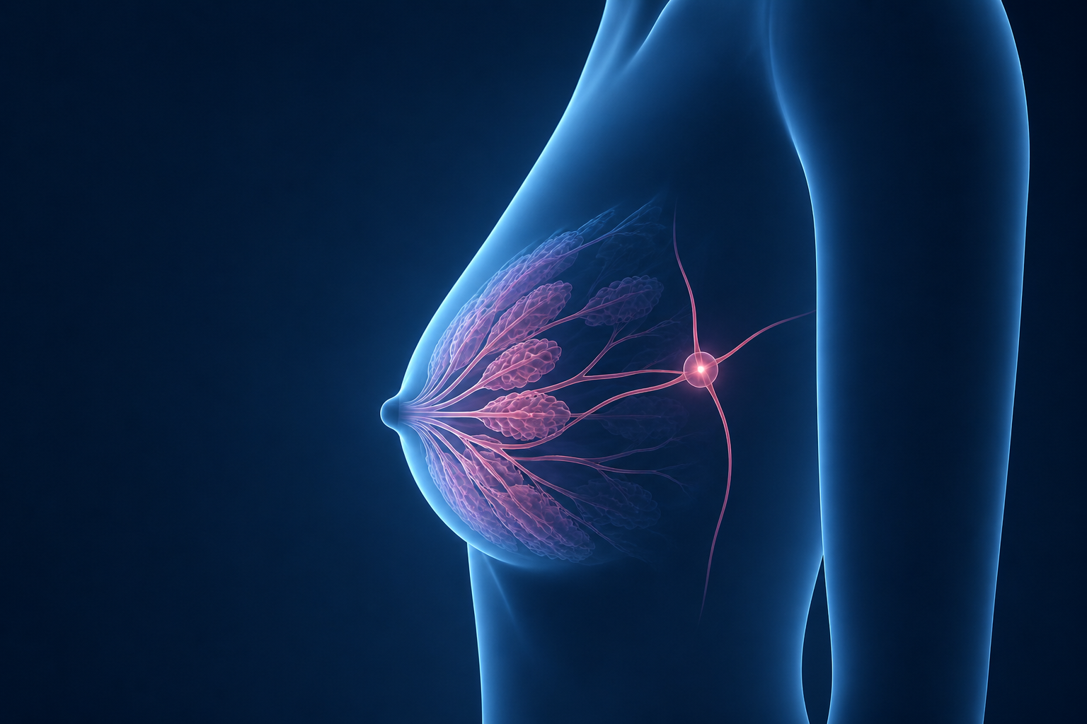
What Exactly Are These Calcifications?
Fine linear branching calcifications are tiny (less than 0.5mm), thin, irregularly shaped calcium deposits within the walls or contents of the breast ducts, arranged in a linear or branching (tree-like) pattern that follows the natural branching anatomy of the duct system.
Why These Calcifications Form
In high-grade DCIS, rapidly growing cancer cells within the ducts die faster than they can be supplied with blood. The dead cell debris (comedo necrosis) undergoes calcification. Because this process follows the duct system, the calcifications follow the ductal branching pattern — creating the characteristic linear or V-shaped and Y-shaped deposits. "Casting calcifications" is another term — the calcifications fill the duct lumen and cast the exact shape of the duct.
What the Radiologist Evaluates
Morphology
Are the individual particles fine and linear (high suspicion) or more amorphous/round (lower suspicion)?
Distribution
Linear (following a single duct) or segmental (triangular, pointing toward the nipple, following an entire ductal segment)? Segmental is most concerning.
Extent
How large an area do the calcifications span? This has direct implications for surgical planning — lumpectomy vs. mastectomy.
Associated Findings
Is there an associated mass? Skin thickening? Axillary lymph node abnormality? These change staging and management.
Types Within This Category
Fine Linear (without branching)
Individual tiny deposits in a linear arrangement. Highly suspicious — BI-RADS 4C or 5.
Fine Linear Branching / Casting (classic BI-RADS 5)
V- or Y-shaped deposits following duct bifurcations. The most suspicious form. Classic presentation of high-grade DCIS.
Fine Pleomorphic in Linear/Segmental Distribution
Varying shapes in a linear or segmental distribution. Also highly suspicious (BI-RADS 4C or 5).
What Happens Next
Multiple cores taken. Specimen radiography confirms the suspicious calcifications were captured in the sample.
Lumpectomy with radiation or mastectomy depending on extent of disease. Sentinel lymph node biopsy may be recommended.
Axillary evaluation, breast MRI, and medical oncology referral begin immediately.
A benign biopsy result cannot be accepted for a BI-RADS 5 finding. Repeat biopsy or surgical excision is required regardless.
Urgent Actions
Act promptly — days, not weeks:
- Biopsy should be arranged urgently
- Bring prior mammograms to your biopsy appointment — comparison helps confirm extent
- Ask about the extent of the calcifications — how large an area they span matters for surgical planning
- Ask whether MRI is recommended before surgery to assess for an invasive component
Related Topics
Does your report mention fine linear calcifications, fine linear branching calcifications, or BI-RADS 5? This is the highest level of suspicion in breast imaging. A board-certified radiologist with subspecialty breast imaging experience can walk you through exactly what it means and what your next steps should be.
Contact the Breast Center